

Hooded eyelids and drooping eyelids are among the most common concerns brought to aesthetic and oculoplastic clinics, yet the two are frequently confused. Understanding what is actually causing your eyelids to droop — whether that is excess skin, a weakened muscle, a descended brow, or an underlying medical condition — is the essential first step before exploring any treatment. This guide explains the distinct causes in plain language, highlights when drooping may signal something that needs urgent medical attention, and outlines the realistic options available for each cause.
Key Takeaways
- Hooded eyelids caused by excess skin (dermatochalasis) and true drooping caused by a weakened eyelid-lifting muscle (blepharoptosis) are different conditions that require different treatments.
- Ageing, genetics, and lifestyle factors are the most common drivers of hooded eyes, but medical conditions including myasthenia gravis, thyroid eye disease, and Horner syndrome can also cause eyelid drooping.
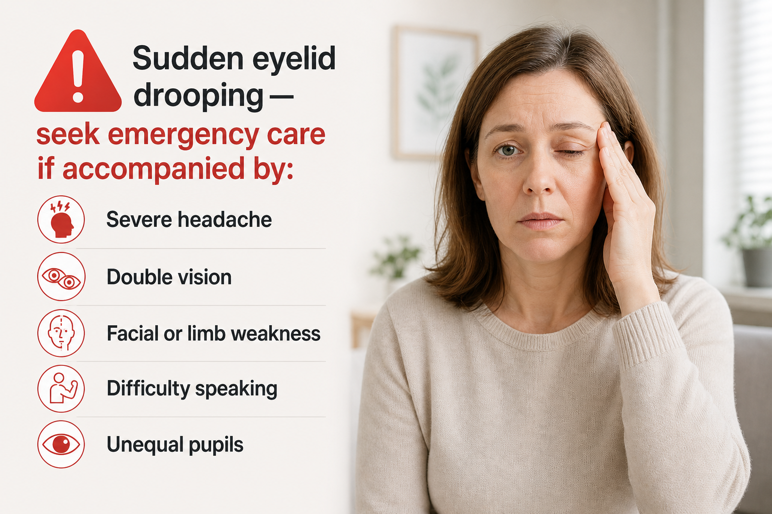
- Sudden-onset drooping of one eyelid — especially with other neurological symptoms — can indicate a stroke or aneurysm and requires immediate emergency assessment.
- Brow ptosis (a descended brow pushing skin over the upper lid) is a distinct and often overlooked cause that changes which treatment is most appropriate.
- Treatment options range from upper blepharoplasty and ptosis repair surgery to non-surgical approaches such as botulinum toxin brow lifts, dermal fillers, and prescription eye drops, depending on the underlying cause.
Hooded Eyes vs. Drooping Eyelids: Understanding the Difference
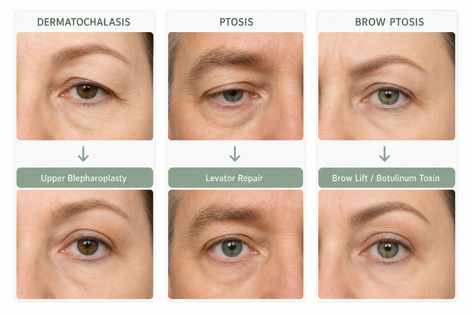
The terms “hooded eyes” and “droopy eyelids” are often used interchangeably, but they describe two clinically distinct situations. Hooded eyes — medically termed dermatochalasis — occur when excess upper eyelid skin folds downward over the eyelid crease, partially obscuring the lid and sometimes the eye itself. The eyelid margin (the edge of the lid closest to the eye) typically sits at a normal height; it is the overlying skin that creates the hooded appearance. Dermatochalasis is primarily a cosmetic and structural issue, though in more advanced cases the overhanging skin can cause visual field obstruction.
True drooping of the eyelid — known medically as blepharoptosis or simply ptosis — is different. In ptosis, the eyelid margin itself sits lower than it should, often partially covering the pupil. This happens because the levator palpebrae superioris, the primary muscle responsible for lifting the upper eyelid, is weakened, stretched, or neurologically compromised. Ptosis can affect one eye (unilateral) or both (bilateral) and ranges from mild to severe.
A third cause — brow ptosis — is frequently overlooked. When the brow descends with age, it pushes the forehead skin and upper eyelid skin downward, creating the appearance of hooded or heavy lids even when the eyelid itself is structurally normal. Many patients have a combination of all three factors simultaneously, which is why accurate diagnosis by a qualified practitioner matters before any treatment is chosen.
| Feature | Dermatochalasis (Hooded Eyes) | Blepharoptosis (Ptosis) | Brow Ptosis |
|---|---|---|---|
| Primary cause | Excess upper eyelid skin | Levator muscle weakness or nerve issue | Descended brow position |
| Eyelid margin position | Normal | Lower than normal | Usually normal |
| Levator function | Intact | Reduced or absent | Intact |
| Common cause | Ageing, genetics, skin laxity | Ageing, neurological, congenital | Ageing, volume loss, gravity |
| Typical treatment | Upper blepharoplasty | Ptosis repair / levator surgery | Brow lift, filler, botulinum toxin |
The Most Common Cause: Ageing and Skin Laxity
Age-related changes are responsible for the majority of hooded eyelid presentations seen in aesthetic practice. From the mid-thirties onward, the skin gradually loses collagen and elastin — the structural proteins that keep it firm and resilient. As collagen and elastin loss accelerates, the upper eyelid skin becomes thinner, less elastic, and increasingly prone to folding downward under the influence of gravity. The result is the characteristic heavy, hooded appearance that many people associate with looking tired or older than they feel.
Alongside skin laxity, the fat pads that sit within and around the orbit (eye socket) shift and redistribute with age. Some areas lose volume, causing hollowing, while others may appear fuller as fat migrates. The brow bone loses soft tissue support, contributing to brow descent. The levator aponeurosis — the tendon connecting the levator muscle to the eyelid — can also stretch or partially detach over decades, lowering the eyelid margin and creating acquired ptosis even in the absence of any neurological problem. This type of age-related ptosis is called aponeurotic ptosis and is one of the most common forms seen in adults.
Genetics and Inherited Anatomy
Genetics play a significant role in determining eyelid shape, skin thickness, and the position of the eyelid crease. Some individuals are born with naturally fuller upper lids, a lower-set eyelid crease, or a greater tendency toward skin laxity — all of which contribute to a hooded appearance from an early age. If hooded eyes run in your family, you are more likely to develop them earlier and more prominently than someone without that genetic predisposition.
Ethnicity also influences eyelid anatomy. In East Asian eyelids, for example, the eyelid crease is often lower or absent due to differences in the attachment of the levator aponeurosis and the distribution of orbital fat. This creates a naturally fuller, less defined upper lid that some describe as hooded. This is a normal anatomical variation, not a medical condition, and any discussion of treatment should be approached with cultural sensitivity and a clear understanding of the individual’s personal goals rather than any external aesthetic standard.
Brow Ptosis: The Often-Missed Third Factor
Brow ptosis — the gradual descent of the eyebrow below its youthful position — is a distinct cause of apparent eyelid heaviness that is frequently attributed to the eyelid itself. As the forehead loses volume and the skin loses elasticity, the brow drops, pushing a fold of forehead and upper eyelid skin over the lid. The body often compensates unconsciously by recruiting the frontalis muscle (the forehead muscle) to hold the brow up, which creates persistent horizontal forehead lines and can cause end-of-day brow fatigue or mild headaches.
Identifying brow ptosis matters clinically because treating the eyelid skin alone — for example, with upper blepharoplasty — may not fully resolve the appearance if the brow remains low. In some cases, a brow lift (endoscopic or open) is the more appropriate primary intervention, or a combination approach addressing both the brow and the eyelid may be recommended. A qualified oculoplastic surgeon or aesthetic practitioner will assess brow position as part of any eyelid evaluation.
Lifestyle Factors That Accelerate Eyelid Drooping
Several lifestyle factors can accelerate the collagen and elastin loss that drives skin laxity around the eyes. UV exposure is one of the most significant: ultraviolet radiation breaks down collagen fibres in the dermis and impairs the skin’s ability to repair itself, speeding up the thinning and loosening of eyelid skin. Consistent use of broad-spectrum SPF around the eye area — using formulations safe for periorbital skin — is one of the most evidence-supported preventive measures available.
Smoking compounds skin ageing through multiple mechanisms, including reduced blood flow to the skin, increased production of enzymes that degrade collagen, and direct oxidative damage. Smokers tend to show more pronounced skin laxity at an earlier age than non-smokers, and this effect is visible around the eyes. Significant weight loss can also contribute: when the face loses substantial volume rapidly, the skin around the eyes may become loose and fold more prominently.
Long-term contact lens use is a less widely known but clinically recognised contributor to acquired ptosis. Repeated manipulation of the upper eyelid during lens insertion and removal can gradually stretch the levator aponeurosis over years, lowering the eyelid margin. Similarly, chronic use of steroid eye drops has been associated with eyelid changes in some patients. These are not reasons to avoid contact lenses or necessary medications, but they are worth discussing with a practitioner if drooping develops.
Medical and Neurological Causes of Drooping Eyelids
When eyelid drooping has a medical rather than cosmetic origin, the underlying condition driving levator muscle weakness must be identified and addressed before any cosmetic intervention is considered. Several systemic and neurological conditions can present with ptosis as a symptom.
Myasthenia Gravis
Myasthenia gravis is an autoimmune condition in which the immune system attacks the junction between nerves and muscles, impairing the ability of muscles — including the levator palpebrae superioris — to contract effectively. Ptosis caused by myasthenia gravis is characteristically variable: it may worsen as the day progresses or after sustained upward gaze, and it can affect one or both eyelids. Double vision (diplopia) is a common accompanying symptom. Myasthenia gravis requires diagnosis and management by a neurologist; cosmetic eyelid procedures are not appropriate until the condition is medically controlled.
Thyroid Eye Disease
Thyroid eye disease (TED), associated with Graves’ disease and other thyroid disorders, causes inflammation and swelling of the tissues behind and around the eye. While TED more commonly causes eyelid retraction (the lid sitting too high), it can also cause ptosis in some presentations, and the associated proptosis (bulging of the eye) and eyelid swelling can create a heavy, drooping appearance. Any eyelid change in a patient with known thyroid disease warrants assessment by an ophthalmologist or endocrinologist.
Horner Syndrome
Horner syndrome results from disruption of the sympathetic nerve pathway that supplies the eye and surrounding structures. It produces a characteristic triad: mild ptosis of the upper eyelid, slight elevation of the lower eyelid (giving the eye a smaller appearance), and a constricted pupil (miosis) — all on the same side. Horner syndrome can be caused by a lung tumour, carotid artery dissection, stroke, or other lesions along the sympathetic chain. New-onset Horner syndrome always requires urgent medical investigation to identify the underlying cause.
Other Medical Causes
Additional medical causes of acquired ptosis include damage to the oculomotor nerve (the third cranial nerve, which also controls several eye movements), which can result from a brain aneurysm, diabetes mellitus, or a tumour. Oculopharyngeal muscular dystrophy — a rare inherited muscle disease — causes progressive ptosis alongside difficulty swallowing. Eyelid tumours, cysts, or neurofibromas can physically weigh down the lid. Prior eye surgery or trauma to the eyelid can also damage the levator mechanism and produce post-surgical ptosis.
Congenital Ptosis: Drooping Eyelids in Children
Congenital ptosis is present from birth and results from incomplete development of the levator palpebrae superioris muscle. It affects one eyelid in most cases. Beyond the cosmetic concern, congenital ptosis carries a meaningful medical risk: if the drooping lid covers the pupil during the critical early years of visual development, it can prevent the eye from receiving adequate visual stimulation, leading to amblyopia (commonly called lazy eye) — a condition in which the brain suppresses input from the affected eye, causing permanent reduction in vision if not treated promptly.
Children with congenital ptosis should be assessed by a paediatric ophthalmologist. The timing of surgical intervention depends on the severity of the ptosis and whether amblyopia is present or at risk of developing. Mild cases may be monitored, while more significant ptosis affecting the visual axis typically requires early surgical correction. Parents who notice one eyelid consistently lower than the other in an infant or young child should seek prompt professional assessment rather than waiting.
When Drooping Is a Medical Emergency
Most cases of hooded or drooping eyelids develop gradually over months or years. However, sudden-onset drooping of one eyelid — particularly when accompanied by other symptoms — can indicate a life-threatening neurological emergency and must be treated as such.
Seek emergency medical care immediately if eyelid drooping appears suddenly alongside any of the following: severe headache (often described as the worst headache of one’s life), double vision, facial weakness or numbness, arm or leg weakness, difficulty speaking, or a change in pupil size. These symptoms may indicate a brain aneurysm, stroke, or carotid artery tear (dissection) — all of which require urgent hospital assessment. A sudden third nerve palsy causing ptosis with a dilated pupil on the same side is a classic warning sign of a posterior communicating artery aneurysm and is a neurological emergency. Do not wait to see if the drooping resolves on its own.
Self-Assessment: Identifying Which Type You May Have
A simple at-home observation can help you understand which type of eyelid concern you may be dealing with, though it does not replace a clinical assessment. Stand in front of a well-lit mirror and look straight ahead at eye level.
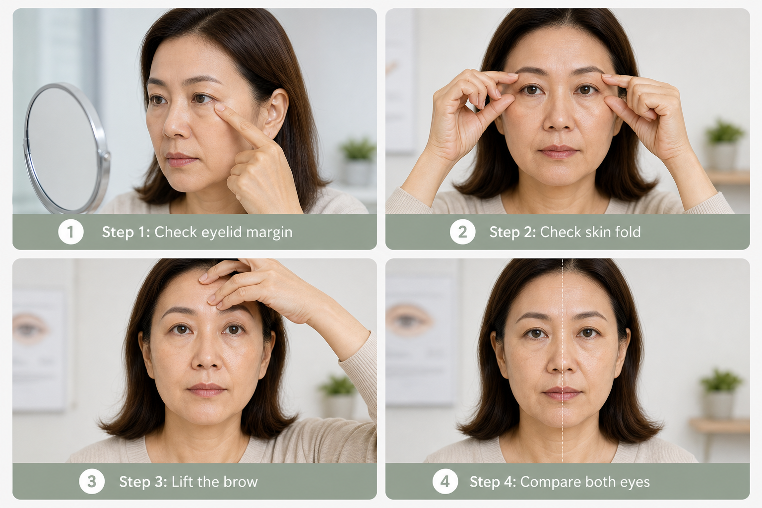
- Check the eyelid margin: The lower edge of your upper eyelid should sit just below the top of the iris (the coloured part of the eye). If the margin is covering a significant portion of the iris or the pupil, this suggests true ptosis rather than simply excess skin.
- Check the skin fold: If the eyelid margin sits at a normal height but a fold of skin hangs over it, obscuring the lid or the eyelid crease, this is more consistent with dermatochalasis (excess skin).
- Check the brow position: Place a finger gently under your brow and lift it slightly to its natural youthful position. If the eyelid heaviness improves significantly when the brow is lifted, brow ptosis may be a contributing factor.
- Check for asymmetry: Compare both eyes. Significant asymmetry — especially if it has appeared recently — warrants professional assessment to rule out a medical cause.
This self-check can help you have a more informed conversation with a practitioner, but the definitive distinction between dermatochalasis, blepharoptosis, and brow ptosis requires examination by a qualified oculoplastic surgeon or experienced aesthetic physician.
Treatment Options: Matching the Solution to the Cause
The appropriate treatment depends entirely on the underlying cause. Applying a cosmetic solution to a medical problem — or treating the wrong anatomical structure — will not produce satisfactory results and may delay necessary care.
Surgical Options
Upper blepharoplasty is the gold-standard surgical treatment for dermatochalasis. The procedure removes carefully measured amounts of excess upper eyelid skin and, where appropriate, repositions or removes herniated orbital fat. It is performed under local anaesthesia in most cases, with a recovery period of approximately one to two weeks for the initial swelling and bruising to settle. Upper blepharoplasty addresses skin laxity effectively but does not correct a low eyelid margin caused by levator muscle weakness — if ptosis is also present, a separate ptosis repair procedure (levator resection or levator advancement) is required at the same time or as a separate operation.
Ptosis repair surgery targets the levator mechanism directly. In levator resection, the levator muscle is shortened to increase its lifting power. In levator advancement, a stretched or detached levator aponeurosis is reattached at a more effective position. Both procedures require precise surgical technique and are best performed by an oculoplastic surgeon with specific training in eyelid anatomy. A brow lift — either endoscopic (minimally invasive) or open — addresses brow ptosis and may be performed alongside eyelid surgery when both are contributing to the appearance.
Non-Surgical Options
Non-surgical approaches can improve mild to moderate hooding and brow descent, though they cannot replicate the results of surgery for significant excess skin or true ptosis. A botulinum toxin brow lift involves carefully placed injections that relax the muscles pulling the brow downward, allowing the frontalis muscle to elevate the brow slightly. Results are temporary, typically lasting three to four months, and the degree of lift is modest — generally most useful for patients with mild brow descent and minimal excess skin.
Dermal fillers placed at the brow bone or temples can restore volume loss that contributes to brow descent, providing subtle structural support. Radiofrequency treatments, laser skin tightening, and ultrasound-based devices such as Ultherapy may offer modest improvement in skin laxity for patients with mild hooding, though results are generally less predictable and less dramatic than surgical correction. For acquired ptosis specifically, oxymetazoline hydrochloride eye drops (available by prescription in some countries under the brand name Upneeq) can temporarily stimulate Müller’s muscle to lift the eyelid margin by approximately 1–2 mm — a useful option for mild acquired ptosis in appropriate candidates. Eyelid tape and ptosis crutches (spectacle-mounted supports) are temporary mechanical aids that some patients use while awaiting surgery or as a long-term non-surgical management strategy.
Frequently Asked Questions
Can hooded eyelids cause vision problems?
Yes, in more advanced cases. When excess upper eyelid skin or a significantly drooping eyelid margin encroaches on the visual field — particularly the superior (upper) field — it can cause visual field obstruction. This is more common with true ptosis than with mild dermatochalasis. When vision is affected, upper blepharoplasty or ptosis repair may be considered medically necessary rather than purely cosmetic, which can affect insurance or funding eligibility in some healthcare systems.
Do hooded eyelids get worse with age?
Generally, yes. The collagen and elastin loss, fat pad redistribution, and brow descent that drive hooded eyelids are progressive age-related processes. Most people notice a gradual worsening over years rather than a sudden change. Lifestyle factors such as UV exposure and smoking can accelerate this progression.
Is sudden eyelid drooping always an emergency?
Sudden-onset drooping — particularly of one eyelid, and especially when accompanied by headache, double vision, facial weakness, or pupil changes — should be treated as a medical emergency until a serious cause is ruled out. Gradual drooping that develops over months or years is far less likely to indicate an acute neurological problem, but any new or unexplained change in eyelid position warrants professional assessment.
Can both dermatochalasis and ptosis occur at the same time?
Yes, and this is clinically common, particularly in older adults. A patient may have both excess upper eyelid skin (dermatochalasis) and a low eyelid margin due to levator muscle weakness (blepharoptosis) simultaneously. When both are present, addressing only one may leave the patient dissatisfied with the outcome. A thorough preoperative assessment should identify both components so that the surgical plan can address each appropriately.
Are there non-surgical ways to prevent hooded eyelids from worsening?
While no non-surgical approach can reverse significant excess skin or true ptosis, consistent sun protection, avoiding smoking, maintaining a stable healthy weight, and using evidence-based skincare ingredients that support collagen production (such as retinoids and vitamin C) may help slow the rate of skin laxity progression around the eyes. These measures are supportive rather than curative.
What is the difference between a brow lift and upper blepharoplasty?
Upper blepharoplasty removes excess skin from the upper eyelid itself. A brow lift elevates the position of the eyebrow, which indirectly reduces the amount of skin pushed over the upper lid. The two procedures address different anatomical structures and are sometimes performed together when both brow descent and excess eyelid skin are contributing to the appearance. Choosing the wrong procedure — for example, performing blepharoplasty alone when brow ptosis is the primary driver — can produce an unsatisfactory result or, in some cases, worsen brow position.
Understanding Your Eyelids Before Choosing a Treatment
Hooded and drooping eyelids have multiple possible causes — excess skin, levator muscle weakness, brow descent, medical conditions, or a combination of these — and the most effective treatment depends on accurately identifying which factor or factors are at play. For the majority of patients, the cause is age-related dermatochalasis, brow ptosis, or both, and a range of surgical and non-surgical options can meaningfully improve the appearance and, where vision is affected, function. For a smaller group, drooping reflects an underlying medical condition that requires diagnosis and management before any cosmetic intervention is appropriate.
If you are concerned about eyelid drooping — whether for cosmetic or functional reasons — a consultation with a qualified oculoplastic surgeon or experienced aesthetic practitioner is the most reliable way to understand what is causing your specific presentation and which treatment options are genuinely suitable for you. Sudden or rapidly worsening drooping, particularly with other neurological symptoms, should prompt immediate emergency assessment rather than a clinic appointment.