

Acrochordons, more commonly known as skin tags, are among the most frequently encountered benign skin growths in adults. They are harmless in the vast majority of cases, yet they often cause concern because of their appearance or because they catch on clothing and jewellery. This article explains what acrochordons are, why they develop, how a clinician distinguishes them from other skin lesions, and what removal options are currently available — including their benefits, limitations, and realistic expectations.
Key Takeaways
- Acrochordons are benign, soft, pedunculated skin growths that most commonly appear in skin fold areas such as the neck, armpits, groin, and eyelids.
- They are not cancerous and do not become cancerous, but any lesion that changes rapidly, bleeds, or looks unusual should be assessed by a qualified clinician before removal.
- Several effective removal methods exist, including cryotherapy, electrocautery, surgical excision, and laser treatment; the most appropriate option depends on lesion size, location, and individual factors.
- At-home removal attempts carry real risks including infection, scarring, and incomplete removal, and are generally not recommended by dermatologists.
- Removing an acrochordon does not prevent new ones from forming, particularly if underlying contributing factors such as insulin resistance or friction remain present.
What Is an Acrochordon?
An acrochordon is a small, soft, flesh-coloured or slightly pigmented growth that attaches to the skin surface via a narrow stalk, known medically as a peduncle. The term comes from the Greek words for extremity and cord, reflecting the characteristic stalk-like connection. Acrochordons are classified as benign fibroepithelial polyps, meaning they consist of loose fibrous connective tissue covered by a layer of normal-appearing skin. They range in size from one or two millimetres — barely visible — to occasionally one or two centimetres in diameter, though larger examples are less common.
Skin tags are extremely prevalent. Estimates suggest that approximately 46% of the general population will develop at least one acrochordon during their lifetime, with incidence increasing significantly after the age of 40. They affect men and women at broadly similar rates, though certain risk factors, discussed below, can increase the likelihood of developing multiple lesions. The growths are almost always asymptomatic unless they are repeatedly irritated by friction, tight clothing, or jewellery, in which case they may become tender, inflamed, or occasionally twist on their stalk and lose their blood supply — a process called torsion.
Where Do Acrochordons Typically Appear?
Acrochordons most commonly develop in areas where skin rubs against skin or clothing. The most frequently affected sites include the neck, particularly along the collar line; the axillae (armpits); the groin and inner thighs; the inframammary fold beneath the breasts; and the eyelids. Less commonly, they may appear on the trunk or face. The distribution pattern strongly suggests that chronic friction and moisture play a role in their formation, which is consistent with their higher prevalence in skin fold areas and in individuals with larger body habitus.
Eyelid acrochordons deserve particular mention because their location near the eye makes self-removal especially inadvisable. Removal in this area requires careful technique by an experienced practitioner to avoid injury to the delicate periorbital structures.
What Causes Skin Tags to Form?
The precise mechanism by which acrochordons develop is not fully established, but several contributing factors are consistently identified in clinical and epidemiological research.
Friction and Mechanical Irritation
Repeated skin-on-skin or skin-on-fabric friction is widely considered a primary mechanical trigger. This explains the characteristic distribution in skin folds and along collar lines. The friction hypothesis is supported by the observation that acrochordons are more common in individuals with obesity, where skin fold contact is more extensive.
Insulin Resistance and Metabolic Factors
A well-documented association exists between multiple acrochordons and insulin resistance, including type 2 diabetes mellitus. Elevated insulin levels are thought to stimulate epidermal and dermal cell proliferation through insulin-like growth factor receptors. Some clinicians regard the presence of numerous skin tags as a potential cutaneous marker of underlying metabolic dysfunction, and a patient presenting with many new acrochordons may benefit from a review of their metabolic health. This does not mean that every person with a skin tag has diabetes — isolated acrochordons are extremely common in otherwise healthy individuals — but the association is clinically relevant when multiple lesions appear.
Hormonal Changes
Acrochordons are more frequently reported during pregnancy, likely due to hormonal shifts and associated weight changes that increase skin fold friction. They may partially regress after delivery in some cases, though this is not universal.
Genetic Predisposition
A familial tendency to develop acrochordons has been observed, suggesting that genetic factors influence individual susceptibility. However, no single causative gene has been identified, and the inheritance pattern appears to be multifactorial.
Human Papillomavirus
Some studies have detected human papillomavirus (HPV) DNA within acrochordon tissue, raising the question of whether certain HPV subtypes contribute to their development. The evidence on this point remains mixed and inconclusive; HPV is not currently considered a confirmed causal factor for acrochordons in the way it is for common warts.
How Are Acrochordons Diagnosed?
In most cases, an experienced clinician can diagnose an acrochordon through visual inspection and dermoscopy alone. The characteristic soft, pedunculated appearance, flesh-coloured or lightly pigmented surface, and typical location are usually sufficient for a confident clinical diagnosis. Dermoscopy — the use of a handheld magnifying device with polarised light — can help confirm the benign nature of the lesion by revealing the characteristic loose connective tissue pattern and absence of features associated with malignancy.
Histological examination (laboratory analysis of a tissue sample) is not routinely required for typical acrochordons. However, if a lesion has an atypical appearance — such as rapid growth, irregular pigmentation, ulceration, or bleeding — a clinician may recommend biopsy or excision with histological analysis to exclude other diagnoses. Conditions that can occasionally resemble acrochordons include seborrhoeic keratoses, neurofibromas, melanocytic naevi (moles), molluscum contagiosum, and, rarely, malignant lesions. This is why professional assessment before any removal attempt is important.
Acrochordon Removal: Available Methods
Acrochordons do not require removal from a medical standpoint, as they are benign and do not pose a health risk. Removal is typically performed for cosmetic reasons or because a lesion is causing persistent irritation. Several clinically established methods are available, each with its own advantages, limitations, and suitability criteria.
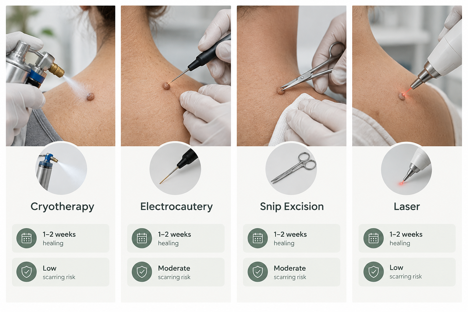
Cryotherapy
Cryotherapy involves applying liquid nitrogen to the acrochordon, causing rapid freezing that destroys the tissue. The treated lesion typically blisters, darkens, and falls away within one to two weeks. Cryotherapy is well-established for small to medium acrochordons and is widely available in dermatology and aesthetic medicine settings. It is generally quick, requires no local anaesthetic for very small lesions, and leaves minimal scarring in most cases. Potential side effects include temporary redness, blistering, post-inflammatory hypopigmentation (lightening of the skin at the treatment site), and, less commonly, hyperpigmentation. Multiple sessions may occasionally be needed for larger lesions.
Electrocautery and Electrodesiccation
Electrocautery uses a controlled electrical current to burn and destroy the acrochordon tissue. Electrodesiccation is a related technique that dehydrates the tissue using high-frequency electrical current. Both methods are effective for acrochordons of varying sizes and can be performed under local anaesthetic. They offer precise control and are particularly useful for lesions in sensitive areas. The treated site heals over one to two weeks, and scarring risk is low when the procedure is performed by an experienced practitioner. A small risk of post-procedural pigmentation change exists, particularly in individuals with darker skin tones.
Surgical Excision and Snip Excision
Snip excision — cutting the acrochordon at its stalk using sterile scissors or a scalpel — is one of the most straightforward and immediately effective removal methods. It is typically performed under local anaesthetic and provides an intact specimen for histological examination if required. Healing is usually rapid for small lesions. Surgical excision is particularly appropriate for larger acrochordons or when tissue analysis is clinically indicated. Minor bleeding at the excision site is common and is usually managed with pressure or a haemostatic agent.
Laser Treatment
Laser removal uses focused light energy to ablate or vaporise acrochordon tissue. Carbon dioxide (CO2) lasers and erbium lasers are among those used for this purpose. Laser treatment offers precision and is particularly useful for multiple lesions or those in cosmetically sensitive areas such as the eyelids or face. It may be associated with a higher cost than other methods and requires appropriate eye protection and practitioner training. Post-treatment care involves keeping the area clean and protected while healing occurs over one to two weeks.
Ligation
Ligation involves tying a fine suture or thread tightly around the stalk of the acrochordon to cut off its blood supply, causing the lesion to necrose and fall away over several days. This technique is sometimes used in clinical settings for pedunculated lesions with a clearly defined stalk. It is not recommended as a self-administered home remedy, as improper technique can lead to infection, incomplete removal, or injury to surrounding skin.
Comparing Removal Methods at a Glance
| Method | Mechanism | Typical Suitability | Healing Time | Scarring Risk | Anaesthetic |
|---|---|---|---|---|---|
| Cryotherapy | Freezing with liquid nitrogen | Small to medium lesions | 1–2 weeks | Low; pigmentation change possible | Usually not required for small lesions |
| Electrocautery | Electrical current burns tissue | Various sizes; sensitive areas | 1–2 weeks | Low with skilled technique | Local anaesthetic common |
| Snip excision | Physical cutting at stalk | Most sizes; histology possible | Days to 1 week | Low for small lesions | Local anaesthetic |
| Laser | Light energy ablation | Multiple or cosmetically sensitive lesions | 1–2 weeks | Low with appropriate settings | Topical or local anaesthetic |
| Ligation | Stalk tied to cut blood supply | Clearly pedunculated lesions | Days to 2 weeks | Low | Usually not required |
Risks and Side Effects of Acrochordon Removal
All removal methods carry some degree of risk, even when performed by a qualified practitioner. Understanding these risks helps patients make an informed decision and set realistic expectations.
- Infection: Any procedure that breaks the skin surface carries a risk of bacterial infection. Signs of infection include increasing redness, warmth, swelling, discharge, or fever, and should prompt prompt medical review.
- Scarring: While scarring risk is generally low for small acrochordons removed with appropriate technique, it cannot be entirely excluded. Individuals with a history of keloid or hypertrophic scarring should discuss this with their clinician before proceeding.
- Pigmentation changes: Post-inflammatory hyperpigmentation or hypopigmentation may occur, particularly with cryotherapy or laser treatment, and is more common in individuals with darker skin phototypes. These changes are often temporary but can occasionally persist.
- Incomplete removal: If the base of the acrochordon is not fully treated, partial regrowth may occur.
- Bleeding: Minor bleeding is expected with excision methods and is usually easily controlled. Patients taking anticoagulant medications should inform their clinician before treatment.
- Recurrence: Removing an acrochordon does not prevent new ones from forming elsewhere, particularly if contributing factors such as friction or metabolic dysfunction persist.
Why At-Home Removal Is Not Recommended
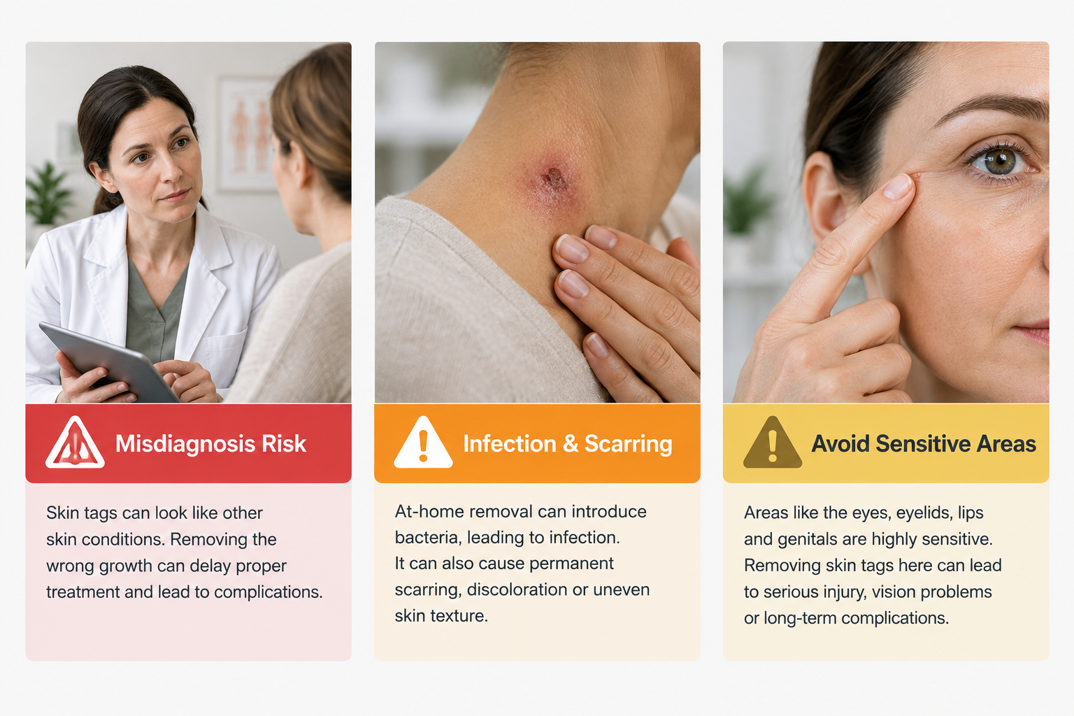
A range of at-home acrochordon removal products and methods are widely available, including over-the-counter freezing kits, ligation bands, and topical preparations. Dermatologists and aesthetic medicine practitioners generally advise against self-removal for several important reasons. First, without professional assessment, there is no certainty that the lesion is a benign acrochordon rather than a different skin condition requiring different management. Second, at-home techniques are difficult to perform with the precision needed to avoid infection, scarring, or incomplete removal. Third, lesions near the eyes, genitals, or other sensitive areas carry particular risks if treated without appropriate training and equipment. If a lesion is causing concern or discomfort, a consultation with a qualified clinician is the safer and more reliable approach.
Who May Be Suitable for Professional Removal?
Most adults with confirmed acrochordons are suitable candidates for professional removal. However, individual assessment is always required. Factors that a clinician will consider include:
- The size, number, and location of the lesions
- The patient’s skin type and history of scarring or pigmentation changes
- Current medications, particularly anticoagulants or immunosuppressants
- Presence of active skin infection or inflammation at or near the treatment site
- Pregnancy, which may affect the timing and choice of treatment
- Any history of bleeding disorders
- Whether the lesion has any atypical features that warrant histological examination
Patients with multiple acrochordons appearing over a short period, or those with associated features such as acanthosis nigricans (a velvety darkening of skin folds), may benefit from a broader metabolic health review alongside any cosmetic treatment discussion.
What to Expect During and After Treatment
The treatment experience varies depending on the method chosen. For most small acrochordons, the procedure is brief — often completed within a few minutes per lesion — and is performed in an outpatient or clinic setting without the need for sedation. Local anaesthetic is commonly used for excision-based methods to minimise discomfort. Cryotherapy may cause a brief stinging or burning sensation during application.
After treatment, the area is typically covered with a small dressing. Patients are usually advised to keep the site clean and dry, avoid picking or scratching the treated area, and apply any prescribed aftercare products as directed. Sun protection over the treated area is important during healing to reduce the risk of pigmentation changes. Most treated sites heal within one to two weeks, though this varies with lesion size, treatment method, and individual healing capacity. A follow-up appointment may be recommended to confirm complete healing and assess the outcome.
Can Acrochordons Be Prevented?
There is no guaranteed method to prevent acrochordons from forming, particularly given the role of genetic predisposition. However, certain practical measures may reduce the likelihood of new lesions developing or existing ones becoming irritated. Maintaining a healthy body weight reduces skin fold friction, which is one of the primary mechanical triggers. Managing blood glucose levels and addressing insulin resistance, where relevant, may also be beneficial given the established metabolic association. Wearing loose-fitting clothing in areas prone to friction and keeping skin folds clean and dry may help reduce irritation. These measures support general skin health but should not be presented as a reliable preventive strategy for acrochordons specifically.
Frequently Asked Questions
Are acrochordons dangerous or a sign of cancer?
Acrochordons are benign growths and are not cancerous, nor do they have any established potential to become cancerous. However, any skin lesion that changes in size, shape, or colour rapidly, bleeds without trauma, or has an unusual appearance should be assessed by a qualified clinician rather than assumed to be a harmless skin tag.
Will an acrochordon go away on its own?
Acrochordons occasionally undergo spontaneous torsion — where the stalk twists, cutting off the blood supply — causing the lesion to darken and fall away on its own. This is not common, and most acrochordons persist indefinitely without treatment. Spontaneous resolution should not be relied upon as a management strategy.
Does removal hurt?
Discomfort during removal depends on the method used and the individual’s pain threshold. Excision-based methods are typically performed under local anaesthetic, making the procedure itself largely painless, though the injection of anaesthetic may cause brief stinging. Cryotherapy may cause a cold, stinging sensation during application. Post-procedure tenderness is usually mild and short-lived.
How many sessions are needed to remove a skin tag?
Most acrochordons can be removed in a single treatment session. Larger lesions or those in particularly sensitive locations may occasionally require a second treatment. Multiple acrochordons can often be treated in the same appointment, depending on their number and distribution.
Will the skin tag grow back after removal?
A correctly and completely removed acrochordon should not regrow at the same site. However, new acrochordons may develop at other locations over time, particularly if the underlying contributing factors — such as friction, metabolic changes, or genetic predisposition — remain present.
Is acrochordon removal covered by health insurance or the NHS?
In most healthcare systems, including the NHS in the United Kingdom, removal of acrochordons for purely cosmetic reasons is not routinely funded. Removal may be considered in cases where a lesion is causing significant functional problems or where there is clinical uncertainty about the diagnosis. Patients seeking removal for cosmetic reasons typically access treatment through private dermatology or aesthetic medicine clinics.
Can I have multiple acrochordons removed at the same appointment?
Yes, in many cases multiple lesions can be treated during a single appointment. The number that can be addressed in one session depends on the treatment method, the size and location of the lesions, and the clinician’s assessment of what is safe and practical for that individual.
Deciding Whether Acrochordon Removal Is Right for You
Acrochordons are a common and benign skin condition that rarely requires treatment from a purely medical perspective. The decision to pursue removal is most often driven by cosmetic preference or by the discomfort caused by repeated irritation. When removal is desired, several safe and effective methods are available through qualified dermatology and aesthetic medicine practitioners, and the choice between them depends on the size, number, and location of the lesions, as well as individual skin characteristics and medical history.
Before proceeding with any removal, a professional assessment is important to confirm the diagnosis, exclude any atypical features that might warrant further investigation, and identify the most appropriate treatment approach. Patients with multiple acrochordons, particularly those appearing over a short period, may also benefit from a broader health review to assess for associated metabolic factors such as insulin resistance.
If you are considering acrochordon removal, a consultation with a qualified dermatologist or experienced aesthetic medicine practitioner is the appropriate first step. A clinician can confirm the diagnosis, discuss the available options in the context of your individual circumstances, and help you understand what to realistically expect from treatment.