

Small red dots on the skin are one of the most common reasons people search for medical information online, and for good reason — they can range from completely harmless to a sign of a serious underlying condition. Understanding what you are looking at, how to perform a simple home assessment, and which symptoms require urgent attention can help you make a more informed decision about whether to wait, see your GP, or go straight to an emergency department.
Key Takeaways
- Pinpoint red dots on the skin are often petechiae — tiny spots caused by bleeding under the skin from broken capillaries — and they do not fade when pressed.
- The glass test (blanch test) is a simple first step: if the spots remain red or purple under a pressed glass, they are non-blanching and need medical assessment.
- Petechiae combined with fever, especially in children, should be treated as a medical emergency until meningococcal disease is ruled out.
- Many red dots, including cherry angiomas and those caused by straining, are benign — but an underlying cause should always be identified when spots appear without explanation.
- Treatment targets the cause, not the spots themselves; petechiae from straining typically resolve within days, while those linked to blood disorders require specialist care.
What Are Petechiae?
Petechiae (pronounced puh-TEE-kee-ee, from the Italian for “small freckle”) are pinpoint-sized, flat red or purple spots that appear when tiny blood vessels called capillaries leak blood into the skin. Because the blood is trapped beneath the surface rather than circulating, the spots do not fade under pressure — a key feature that distinguishes them from most rashes and inflammatory skin conditions. Each individual spot is typically less than 2 mm in diameter, and they usually appear in clusters rather than as isolated marks.
Petechiae are not a rash in the conventional sense. They are not raised, not itchy, and not caused by inflammation or an allergic reaction. They are the visible result of bleeding into the skin from broken capillaries, which can happen for reasons ranging from a bout of heavy coughing to a serious blood disorder. This distinction matters because it changes both the urgency of assessment and the type of treatment required.
When spots are larger than 2 mm but share the same non-blanching, flat appearance, they are classified as purpura. Both petechiae and purpura represent bleeding under the skin; the size difference is simply a clinical convention used to guide further investigation.
How to Perform the Glass Test at Home
The glass test — also called the blanch test — is a straightforward way to assess whether red spots on the skin are non-blanching. It does not replace a medical examination, but it provides useful information before you contact a healthcare professional.
- Find a clear drinking glass. A transparent glass or tumbler works well; you need to be able to see the skin through it.
- Press the glass firmly against the affected area. Apply steady, even pressure so the skin flattens visibly beneath the glass.
- Observe the spots while the glass is pressed. Look carefully at the colour of the dots through the glass.
- Interpret the result. If the spots fade or disappear under pressure, they are blanching — consistent with a standard rash, inflammation, or a cherry angioma. If the spots remain red or purple and do not fade, they are non-blanching petechiae or purpura, and medical assessment is needed.
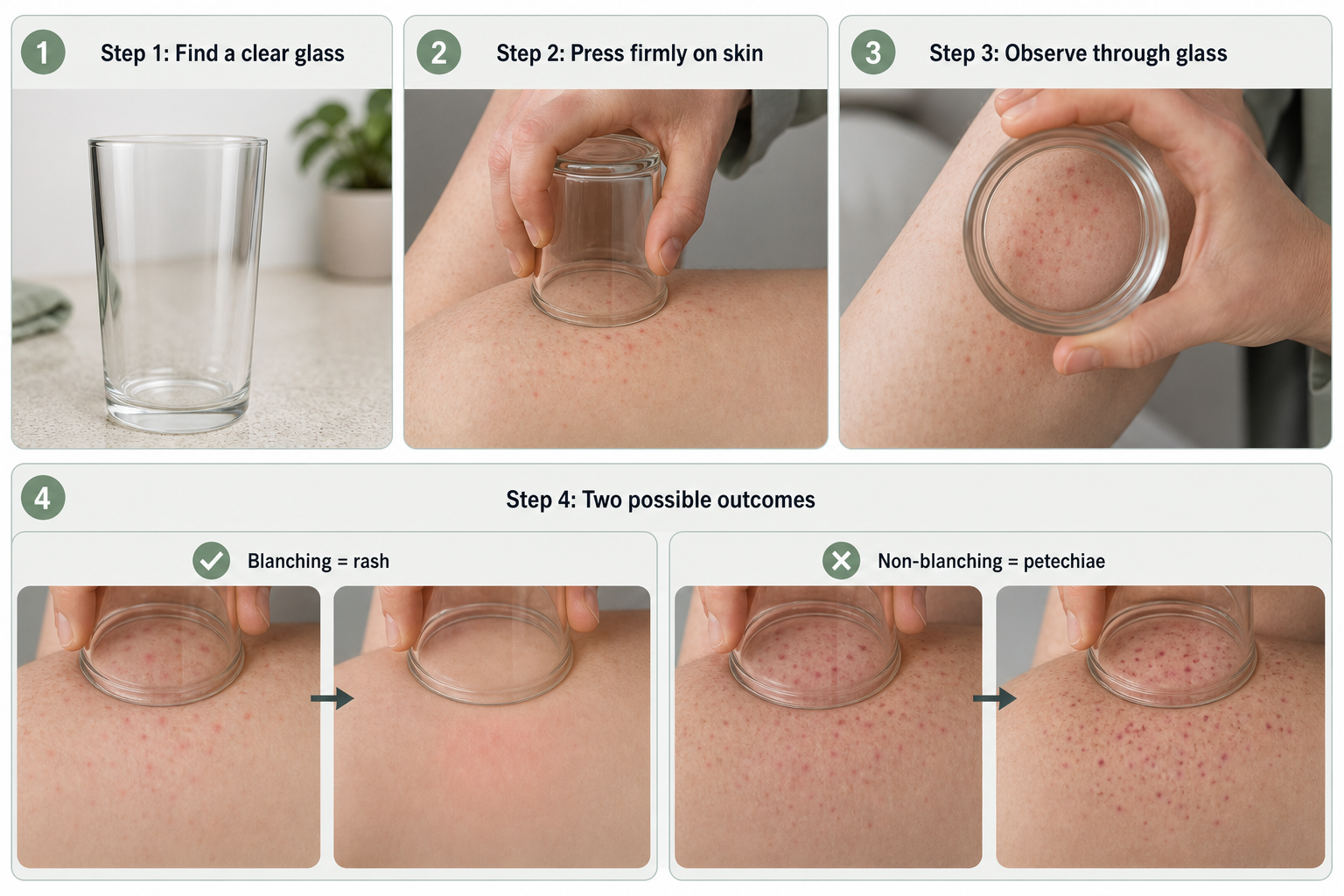
On darker skin tones, petechiae may appear brown or dark purple rather than bright red, and they can be harder to see against the surrounding skin. In these cases, checking areas where the skin is thinner — such as the inner forearm, the palms, the soles, or the mucous membranes inside the mouth — may make the spots easier to identify. Palatal petechiae, which are spots on the roof of the mouth, are sometimes the first visible sign of strep throat or infectious mononucleosis.
Common Causes of Red Dots on Skin
The underlying cause of petechiae spans a wide spectrum of severity. Identifying the most likely cause depends on where the spots appear, whether they are accompanied by other symptoms, and whether they appeared after a specific event such as illness or physical strain.
Mechanical and Benign Causes
Prolonged straining is one of the most common and least concerning causes of petechiae. Intense coughing, vomiting, weightlifting, or the physical effort of childbirth can temporarily raise pressure in small blood vessels enough to cause capillary bleeding. In these cases, the spots typically appear on the face, around the eyes, or on the upper chest — generally above the nipple line — and resolve within a few days without treatment. Physical trauma, friction, or sunburn can produce similar localised spots.
Infections
Viral infections, including the common cold, strep throat, and COVID-19, can cause petechiae through a combination of capillary fragility, immune activation, and, in some cases, mild platelet changes. These spots are usually self-limiting and resolve as the infection clears. However, certain bacterial infections — particularly meningococcal disease — can produce a rapidly spreading non-blanching rash that represents a genuine medical emergency. Endocarditis (infection of the heart valves) and septicaemia are other serious infections that may cause petechiae as part of a broader clinical picture.
Blood Disorders and Low Platelet Count
Platelets are the blood cells responsible for clotting. When the platelet count falls — a condition called thrombocytopenia — even minor capillary stress can result in bleeding under the skin. Immune thrombocytopenia (ITP) is an autoimmune condition in which the immune system destroys platelets, leading to petechiae, easy bruising, and sometimes bleeding from the gums or nose. Leukaemia, lymphoma, and other blood cancers can also suppress platelet production, making unexplained petechiae — particularly when accompanied by fatigue, weight loss, or recurrent infections — a reason for prompt investigation. A complete blood count (CBC) is the standard first-line laboratory test used to assess platelet levels and identify blood disorders.
Autoimmune and Inflammatory Conditions
Vasculitis — inflammation of the blood vessel walls — can cause non-blanching spots by damaging capillaries directly. Henoch-Schönlein purpura (HSP), now more formally called IgA vasculitis, is a form of vasculitis that most commonly affects children and produces a distinctive purpuric rash on the legs and buttocks, often alongside joint pain and abdominal discomfort. Lupus and other systemic autoimmune conditions may also cause petechiae as part of their broader effects on blood vessels and platelets.
Medications and Nutritional Deficiencies
Certain medications can cause petechiae either by reducing platelet count or by increasing capillary fragility. Anticoagulants, some antibiotics (including penicillin), phenytoin, and quinine are among the drugs associated with drug-induced petechiae. Scurvy — caused by severe vitamin C deficiency — is a less common but important cause; vitamin C is essential for collagen synthesis, and without it, capillary walls become fragile and prone to bleeding. Malnutrition more broadly can contribute to capillary fragility and impaired platelet function.
Pigmented Purpuric Dermatoses (Schamberg Disease)
Pigmented purpuric dermatoses, of which Schamberg disease is the most common type, represent a group of chronic skin conditions characterised by clusters of tiny red-brown spots with a distinctive “cayenne pepper” appearance. The spots result from slow leakage of red blood cells from capillaries into the skin, typically on the lower legs. Unlike petechiae from acute causes, Schamberg disease tends to be persistent and recurrent, though it is generally benign and not associated with systemic illness. It is significantly underrepresented in consumer health resources, and many people with this pattern of spots are unaware of the diagnosis.
Petechiae vs. Other Red Dots: A Comparison
| Condition | Size | Appearance | Blanches? | Itchy? | Typical Cause | Urgency |
|---|---|---|---|---|---|---|
| Petechiae | Under 2 mm | Flat, red or purple, pinpoint | No | No | Capillary bleeding; many causes | Varies — assess promptly |
| Purpura | Over 2 mm | Flat, red, purple or brown | No | No | Larger capillary bleed; vasculitis | Medical assessment needed |
| Cherry angioma | 1–5 mm | Raised, bright red, smooth dome | Yes (partially) | No | Benign overgrowth of blood vessels | Low — benign |
| Spider angioma | Central dot with radiating vessels | Red centre, spider-leg pattern | Yes | No | Dilated capillaries; liver disease, pregnancy | Low to moderate |
| Heat rash (miliaria rubra) | 1–4 mm | Small red bumps, may blister | Yes | Yes | Blocked sweat ducts in heat | Low — self-limiting |
| Folliculitis | Variable | Red, hair-centred, may have pus | Yes | Sometimes | Bacterial or fungal follicle infection | Low to moderate |
| Keratosis pilaris | 1–2 mm | Rough, sandpaper-like, skin-coloured or red | Yes | Rarely | Keratin plugs in hair follicles | Low — cosmetic concern |
| Schamberg disease | Pinpoint clusters | Cayenne pepper pattern, brown-red | No | Rarely | Chronic capillary leakage | Low — chronic but benign |
Cherry angiomas deserve particular mention because they are extremely common — especially in adults over 30 — and are frequently mistaken for petechiae. A cherry angioma is a raised, bright red dome caused by a benign overgrowth of blood vessels in the skin. Unlike petechiae, cherry angiomas are slightly raised to the touch, tend to blanch at least partially under pressure, and do not appear suddenly in clusters. They are harmless and require no treatment unless they bleed or cause cosmetic concern, in which case laser treatment or electrocautery can remove them effectively.
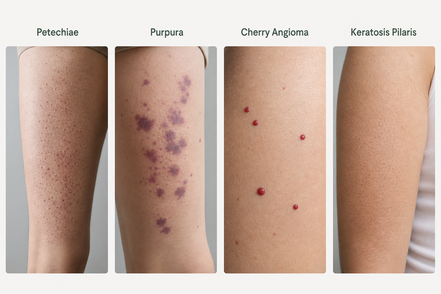
Keratosis pilaris is another condition that generates concern but is entirely benign. It produces rough, sandpaper-like bumps — often with a reddish tinge — on the upper arms, thighs, and cheeks. These bumps are caused by keratin plugging hair follicles and are not itchy, not raised in the same way as a welt, and not related to capillary bleeding. Keratosis pilaris is a cosmetic concern rather than a medical one.
Where Red Dots Appear and What Location May Suggest
The location of petechiae on the body can offer useful clues about their likely cause. Spots confined to the face, around the eyes, or on the upper chest — particularly above the nipple line — after a bout of coughing, vomiting, or heavy lifting are strongly suggestive of mechanical straining and are generally benign. Spots that appear on the lower legs in a chronic, recurring pattern are more consistent with Schamberg disease or venous insufficiency. A widespread distribution across the trunk and limbs, especially when accompanied by systemic symptoms, raises concern for a blood disorder, vasculitis, or serious infection.
Petechiae on the mucous membranes — including the inside of the cheeks, the gums, or the roof of the mouth (palatal petechiae) — can indicate strep throat, infectious mononucleosis, or, less commonly, a platelet disorder. Spots appearing on the eyelids or the white of the eye (conjunctival petechiae) may follow intense straining but can also be associated with endocarditis. Any petechiae that appear on the palms or soles warrant prompt medical review, as this distribution is less typical of benign mechanical causes.
Petechiae in Children: A Higher-Stakes Assessment
In children, the combination of fever with petechiae must be treated as a potential medical emergency until meningococcal disease has been excluded. Meningococcal bacteria can cause a rapidly progressive infection that produces a characteristic non-blanching rash — initially petechiae that may quickly enlarge into purpura — alongside fever, stiff neck, sensitivity to light, and deteriorating consciousness. The speed at which this condition can progress means that a febrile child with non-blanching spots should receive urgent clinical assessment, typically in an emergency department, rather than a wait-and-see approach.
Viral infections are the most common cause of petechiae in children overall, and the majority of cases in a well-appearing child without fever will have a benign explanation. However, because the consequences of missing meningococcal disease are severe, clinical guidelines in many countries recommend that any child with fever and a non-blanching rash be assessed urgently. Parents who are uncertain should always err on the side of seeking immediate medical attention.
When to Seek Medical Attention
Not every case of petechiae requires emergency care, but certain combinations of symptoms should prompt immediate action. The following situations warrant calling emergency services or going directly to an emergency department:
- Petechiae or purpura appearing alongside fever, especially in a child
- Spots that are spreading quickly over minutes or hours
- Accompanying symptoms of confusion, stiff neck, severe headache, or sensitivity to light
- Difficulty breathing, dizziness, or signs of shock
- Petechiae in a person who is already known to have a blood disorder or is taking anticoagulant medication
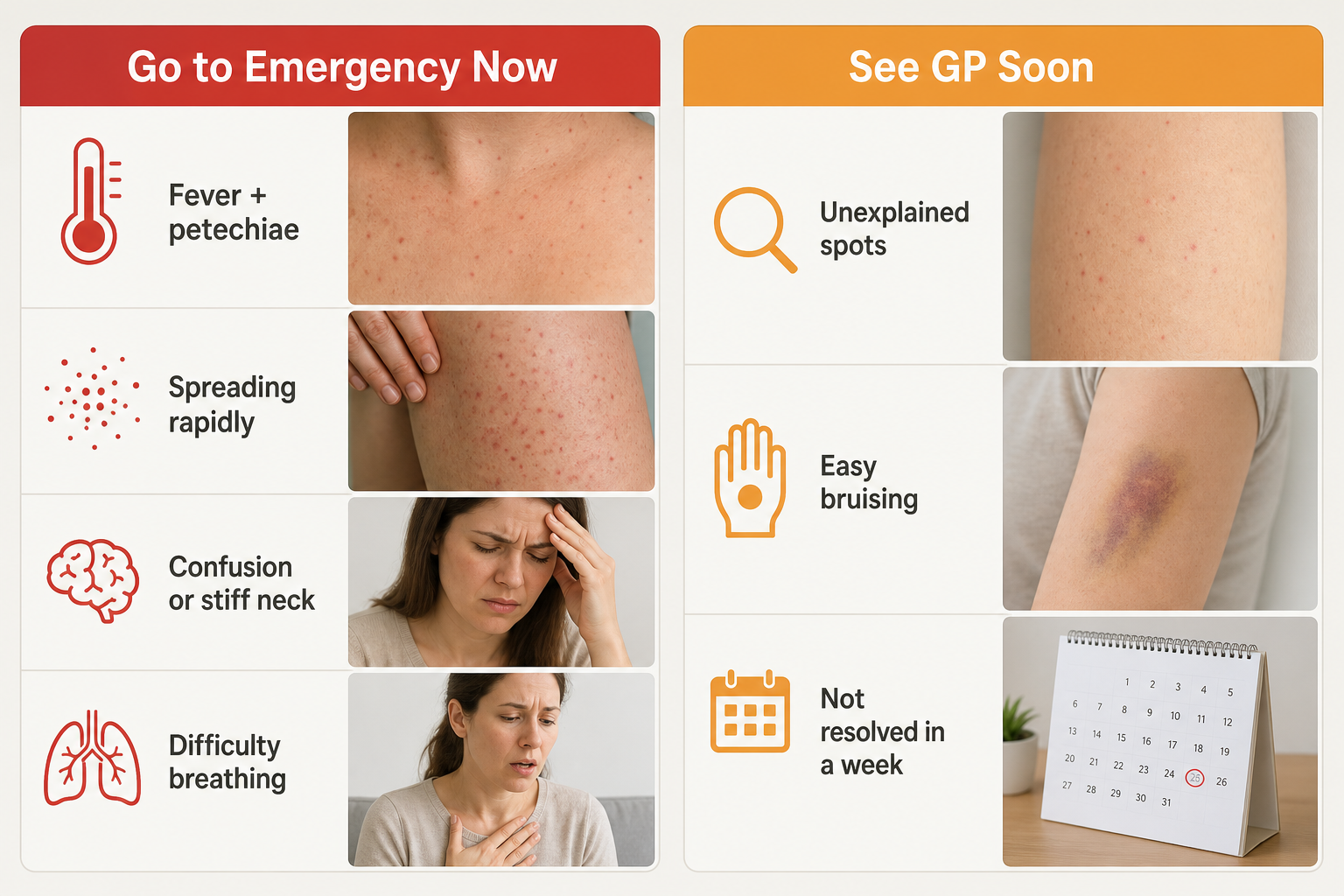
The following situations warrant a same-day or next-day GP appointment rather than emergency care:
- Petechiae that appeared without an obvious mechanical cause such as coughing or straining
- Spots accompanied by easy bruising, bleeding gums, or frequent nosebleeds
- Petechiae that have not resolved within a week
- Recurring episodes of unexplained petechiae
- Spots appearing alongside unexplained fatigue, weight loss, or swollen lymph nodes
Petechiae that appeared directly after a clear mechanical event — such as a prolonged coughing fit or heavy vomiting — in an otherwise well adult, and that are confined to the face or upper chest, can generally be monitored at home. If they do not resolve within a few days, or if new symptoms develop, medical review is appropriate.
How Long Do Petechiae Last?
The duration of petechiae depends almost entirely on their underlying cause. Spots caused by mechanical straining typically fade within two to five days as the leaked blood is reabsorbed by surrounding tissue. The colour may shift from red to brown or purple before disappearing, which is a normal part of the healing process and does not indicate worsening.
Petechiae linked to a viral infection usually resolve alongside the infection itself, typically within one to two weeks. Those caused by a low platelet count — whether from immune thrombocytopenia, medication, or a blood disorder — will persist and may continue to appear until the platelet count recovers. In leukaemia-related cases, petechiae evolve from red to brown and do not fully resolve until platelet production is restored through treatment. Schamberg disease produces spots that may persist for months or years, fading and recurring in a chronic pattern.
Diagnosis and Treatment
A doctor assessing petechiae will typically begin with a detailed history — asking about recent illness, medications, straining events, and associated symptoms — followed by a physical examination. The glass test is often performed clinically to confirm the non-blanching nature of the spots. A complete blood count (CBC) is the standard first-line blood test, providing information about platelet levels, red blood cell count, and white blood cell count. Depending on the results, further tests may include blood clotting studies, inflammatory markers, blood cultures, or urine analysis.
Treatment is directed at the underlying cause rather than the petechiae themselves. Straining-related petechiae require no specific treatment. Bacterial infections are treated with antibiotics; meningococcal disease requires intravenous antibiotics in hospital. Immune thrombocytopenia may be managed with corticosteroids, intravenous immunoglobulin, or other immunosuppressive therapies. Blood cancers such as leukaemia require specialist oncology care, which may include chemotherapy, targeted therapy, or stem cell transplantation. A referral to a haematologist or oncologist is appropriate when a blood disorder or malignancy is suspected. Vasculitis is managed according to its specific type and severity, often involving immunosuppressive medication.
There is no topical treatment that removes petechiae directly. Cosmetic procedures such as laser therapy are occasionally used for persistent cherry angiomas or spider angiomas once a benign diagnosis has been confirmed, but they have no role in the management of true petechiae.
Can Petechiae Be Prevented?
Prevention depends on the cause. Meningococcal vaccination significantly reduces the risk of meningococcal disease and its associated petechiae and purpura; vaccination schedules vary by country and age group, and it is worth confirming your vaccination status with your GP. Avoiding unnecessary straining — for example, by treating chronic cough or constipation — can reduce the frequency of mechanical petechiae. People taking medications known to affect platelet function or capillary fragility should discuss the risk with their prescribing doctor rather than stopping medication independently. Maintaining adequate vitamin C intake through diet or supplementation prevents scurvy-related capillary fragility, though true scurvy is uncommon in countries with access to a varied diet.
Frequently Asked Questions
Are petechiae dangerous?
Petechiae themselves are not dangerous, but they can be a sign of a serious underlying condition. The spots are a symptom, not a disease. Whether they are dangerous depends entirely on what is causing them. Petechiae from straining are harmless; petechiae from meningococcal disease or leukaemia require urgent treatment. This is why identifying the cause matters more than the spots themselves.
Can petechiae go away on their own?
Yes, in many cases. Petechiae caused by mechanical straining or a mild viral infection typically resolve within a few days to two weeks without any specific treatment. Petechiae linked to an ongoing medical condition — such as a low platelet count or vasculitis — will not resolve until the underlying condition is treated.
What do petechiae look like on darker skin tones?
On darker skin tones, petechiae may appear brown, dark purple, or be difficult to see against the surrounding skin. Checking areas with thinner or lighter skin — such as the inner forearm, palms, soles, or the inside of the mouth — can make them easier to identify. The glass test works the same way regardless of skin tone.
Is a cherry angioma the same as petechiae?
No. A cherry angioma is a raised, bright red dome caused by a benign overgrowth of blood vessels. It blanches at least partially under pressure and is not caused by bleeding under the skin. Petechiae are flat, do not blanch, and result from capillary bleeding. Cherry angiomas are harmless and very common in adults over 30.
What is the difference between petechiae and purpura?
Both petechiae and purpura are non-blanching spots caused by bleeding under the skin. The distinction is size: petechiae are less than 2 mm in diameter, while purpura are larger than 2 mm. Both require medical assessment to identify the underlying cause, and purpura — particularly in a febrile child — is treated with greater urgency.
Should I go to A&E for petechiae?
Go to an emergency department immediately if petechiae are accompanied by fever (especially in a child), are spreading rapidly, or are associated with confusion, stiff neck, severe headache, or difficulty breathing. If the spots appeared after straining, are confined to the face or upper chest, and you feel otherwise well, a GP appointment is more appropriate than emergency care — though you should seek that appointment promptly if the spots do not resolve within a few days.
When Red Dots on Skin Need More Than a Wait-and-See Approach
Most red dots on the skin have a straightforward explanation, and many resolve without any intervention. The glass test is a genuinely useful first step — if the spots blanch, they are almost certainly not petechiae, and the urgency is lower. If they do not blanch, the next question is whether there are accompanying symptoms that change the picture. Fever with petechiae, rapidly spreading spots, or systemic symptoms such as confusion or difficulty breathing all shift the assessment firmly toward urgent care.
For spots that are non-blanching but appear in a well person after an obvious mechanical event, a GP review within a day or two is a reasonable and responsible approach. For unexplained petechiae — particularly those accompanied by easy bruising, bleeding from the gums, or unusual fatigue — a complete blood count and clinical assessment should not be delayed. Identifying the underlying cause is the only way to determine whether reassurance, monitoring, or active treatment is appropriate. If you are uncertain about what you are seeing, seeking professional advice is always the right decision.