

Breast dimpling is a change in the skin surface of the breast that can sometimes resemble the texture of an orange peel or a subtle indentation. While it does not always indicate something serious, breast dimpling is recognised as one of the visual warning signs that warrants prompt medical evaluation, because in some cases it may be associated with underlying breast cancer. Understanding what breast dimpling looks like, why it occurs, and when to seek assessment can make a meaningful difference to outcomes.
Key Takeaways
- Breast dimpling refers to a puckering, indentation, or orange-peel texture on the skin of the breast and should always be assessed by a qualified healthcare professional.
- It can be caused by benign conditions, but it is also a recognised warning sign of certain types of breast cancer, including inflammatory breast cancer.
- Early medical assessment is strongly recommended — do not wait to see if the change resolves on its own.
- Breast dimpling is best identified through regular self-examination combined with routine clinical breast screening.
- Other accompanying changes, such as nipple inversion, skin redness, or a new lump, increase the urgency of seeking medical review.
What Is Breast Dimpling?
Breast dimpling describes a visible change in the skin of the breast where the surface appears to pucker, indent, or take on an uneven texture. The affected area may look as though the skin is being pulled inward from beneath, or it may develop a roughened, pitted appearance sometimes described as peau d’orange — a French term meaning “skin of an orange.” This texture occurs when the small pores around hair follicles become more prominent due to swelling or tethering of the underlying tissue.
The dimpling may affect a small localised area or a larger portion of the breast. It can appear on any part of the breast surface, including near the nipple or areola, on the lower pole, or toward the outer edges. In some cases the change is subtle and only visible when the arms are raised or when leaning forward. This is why clinical breast examination often involves observing the breast from multiple positions and angles.
Breast dimpling is not a diagnosis in itself — it is a physical sign that requires investigation to determine its underlying cause. Some causes are benign, but because breast dimpling can be associated with malignant conditions, it should never be dismissed without professional assessment.
Why Does Breast Dimpling Occur?
The breast contains a network of ligaments called Cooper’s ligaments, which connect the skin to the deeper breast tissue and chest wall. These ligaments help maintain the shape and position of the breast. When something disrupts or tethers these ligaments — whether a tumour, inflammation, scar tissue, or another structural change — the overlying skin can be pulled inward, creating the dimpled or puckered appearance.
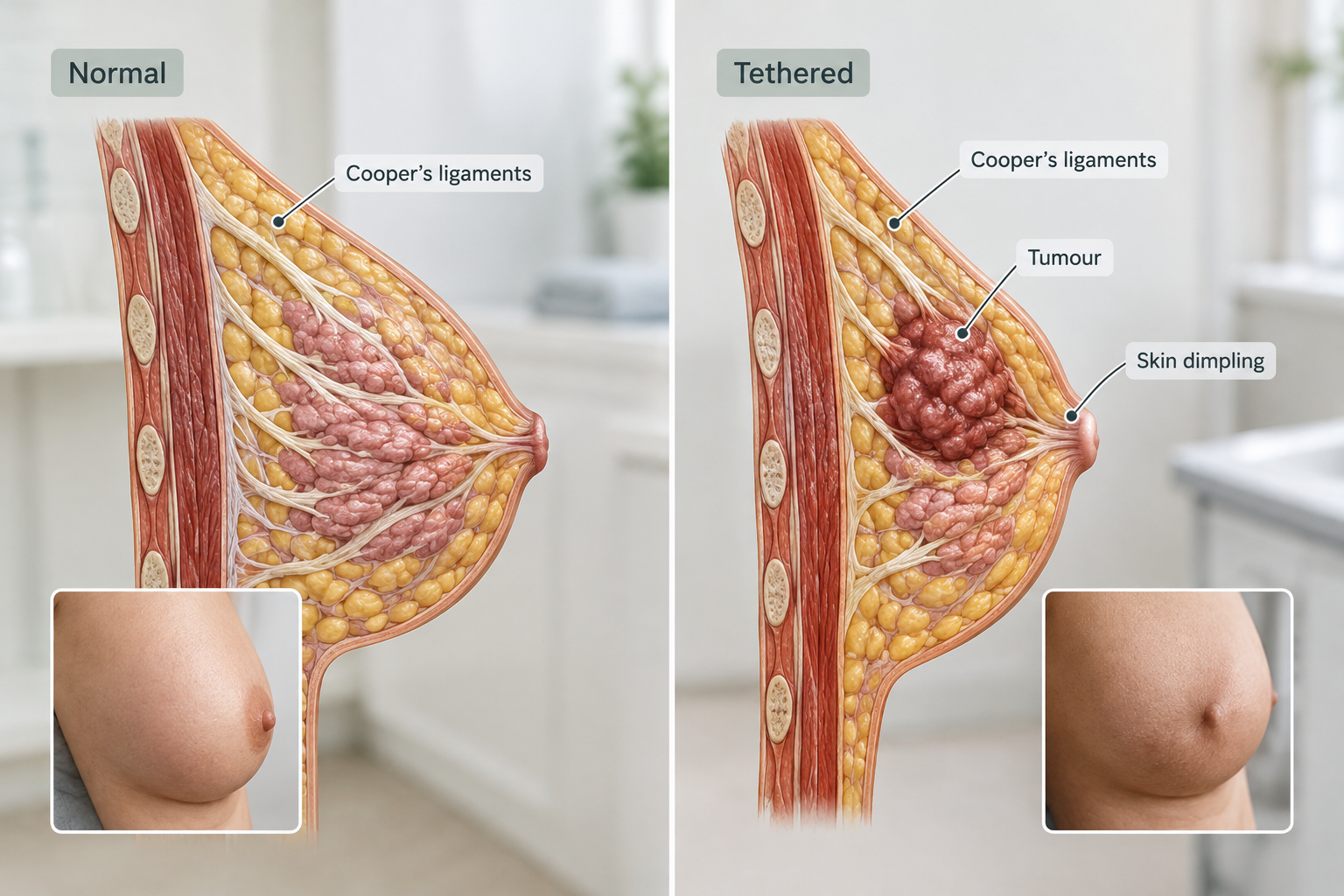
In the context of breast cancer, a tumour growing within the breast tissue may directly invade or compress Cooper’s ligaments, causing them to shorten and tether the skin above. This tethering effect produces the characteristic dimpling or skin retraction associated with certain breast cancers. The same mechanism can cause nipple retraction when the tumour is located near the nipple-areola complex.
In inflammatory breast cancer — a less common but aggressive form of the disease — cancer cells block the lymphatic vessels within the skin of the breast. This blockage causes fluid to accumulate, leading to swelling, redness, warmth, and the distinctive peau d’orange texture. Inflammatory breast cancer may not always present with a palpable lump, which makes skin changes such as dimpling particularly important to recognise.
Not all causes of breast dimpling are malignant. Benign conditions including fat necrosis (damage to fatty tissue, often following trauma or surgery), a benign breast cyst, mastitis (breast infection), or previous surgical scarring can also produce skin tethering and dimpling. However, because the appearance of benign and malignant causes can overlap significantly, clinical and imaging assessment is always required to distinguish between them.
Breast Dimpling as a Warning Sign of Breast Cancer
Breast dimpling is listed by major cancer organisations, including Cancer Research UK and the NHS, as one of the key visual warning signs of breast cancer. It is particularly associated with locally advanced breast cancer and inflammatory breast cancer, though it can occur with earlier-stage disease when a tumour is positioned close to the skin or Cooper’s ligaments.
Inflammatory breast cancer deserves specific attention because it can progress rapidly and may not present with the lump that many people associate with breast cancer. Instead, the breast may appear swollen, feel warm or tender, look red or bruised, and develop the peau d’orange texture across a significant portion of the skin. These changes can sometimes be mistaken for a breast infection, which may delay diagnosis. If antibiotic treatment for a presumed infection does not produce clear improvement within one to two weeks, further investigation for inflammatory breast cancer is essential.
Breast dimpling associated with cancer may appear alongside other warning signs, including a new or changing lump within the breast or armpit, nipple inversion or discharge, changes in breast size or shape, skin thickening, persistent breast pain, or visible veins on the breast surface. The presence of multiple changes together increases the clinical concern and the urgency of assessment.
It is important to understand that breast dimpling does not confirm a diagnosis of cancer. Many people who notice dimpling will have a benign explanation identified after investigation. However, the only way to determine the cause with confidence is through professional clinical examination and appropriate imaging.
How to Check for Breast Dimpling
Regular breast self-examination helps individuals become familiar with the normal look and feel of their breasts, making it easier to notice changes. Breast awareness does not require a rigid technique, but a consistent approach improves the likelihood of detecting changes early.
To check for breast dimpling, stand in front of a mirror with good lighting. Begin with arms relaxed at the sides and observe both breasts for any visible changes in shape, contour, or skin texture. Then raise both arms above the head and look again — this position can reveal dimpling or skin tethering that is not visible when the arms are down. Leaning forward slightly can also help make subtle skin changes more apparent. Finally, place hands on hips and press firmly to contract the chest muscles, which can highlight asymmetry or skin changes.
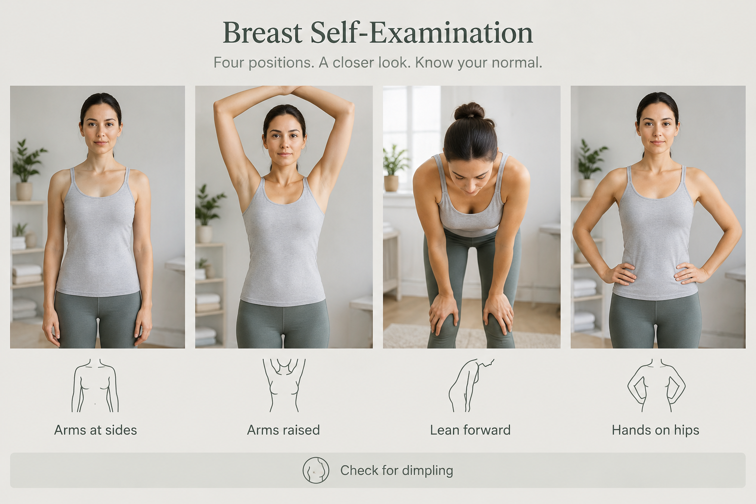
During visual inspection, look specifically for:
- Any area of skin that appears puckered, indented, or pulled inward
- A roughened or orange-peel texture on any part of the breast
- Changes in the position or shape of the nipple
- Redness, swelling, or warmth that is new or unexplained
- Any visible lump or change in breast contour
- Differences between the two breasts that were not previously present
Self-examination should be performed regularly — many clinicians suggest monthly checks — and ideally at the same point in the menstrual cycle, as hormonal changes can cause normal variation in breast texture and size. Self-examination is a useful tool for awareness but is not a substitute for clinical breast screening or mammography.
When to Seek Medical Assessment
Any new or unexplained breast dimpling should be assessed by a doctor promptly. This applies regardless of whether the dimpling is accompanied by other symptoms. The NHS and major breast cancer charities consistently advise that people should not wait to see if a breast change resolves on its own before seeking medical advice.
Seek urgent medical assessment if breast dimpling is accompanied by any of the following:
- A new lump or thickening in the breast or armpit
- Nipple inversion, discharge, or crusting
- Redness, warmth, or swelling of the breast skin
- Rapid change in breast size or shape
- Persistent breast pain that is new and unexplained
- Skin that appears thickened or feels different to the touch
A general practitioner will typically perform an initial clinical examination and, if concerned, refer the patient to a specialist breast clinic. In the UK, an urgent referral under the two-week wait pathway is available for people with symptoms that may indicate breast cancer. At the breast clinic, assessment usually involves a combination of clinical examination, imaging (mammography and/or ultrasound), and in some cases a biopsy to obtain a tissue sample for laboratory analysis.
Early assessment does not mean that cancer will be found — the majority of breast changes investigated at specialist clinics turn out to have benign causes. However, when cancer is present, earlier detection is associated with a wider range of treatment options and generally better outcomes.
Diagnostic Investigations for Breast Dimpling
When a clinician identifies or is told about breast dimpling, the investigation pathway typically involves several steps. The specific investigations recommended will depend on the individual’s age, personal and family history, and the clinical findings at examination.
Clinical breast examination involves a trained clinician assessing the breast visually and by palpation (feeling the tissue). The clinician will look for skin changes, assess the texture and mobility of any palpable abnormality, and examine the lymph nodes in the armpit and around the collarbone.
Mammography is an X-ray of the breast tissue and is the primary imaging tool for breast cancer detection in women over 40. It can identify masses, areas of calcification, and architectural distortion within the breast tissue that may not be palpable. In younger women, breast tissue tends to be denser, which can limit mammographic sensitivity.
Breast ultrasound uses sound waves to produce images of breast tissue and is particularly useful in younger women with denser breasts, for evaluating a specific area of concern, and for distinguishing between solid masses and fluid-filled cysts. Ultrasound is often used alongside mammography rather than as a replacement.
MRI of the breast may be recommended in specific circumstances, such as for individuals with a high genetic risk of breast cancer, when other imaging is inconclusive, or to assess the extent of disease when cancer has been identified.
Core needle biopsy involves removing a small sample of breast tissue using a needle, usually guided by ultrasound or mammography. The sample is examined under a microscope by a pathologist to determine whether cancer cells are present and, if so, what type of cancer it is. A biopsy is the only way to confirm a diagnosis of breast cancer.
Benign Causes of Breast Dimpling
While breast dimpling requires investigation, it is worth understanding that several non-cancerous conditions can produce a similar appearance. Recognising these possibilities does not reduce the importance of seeking assessment, but it may help to contextualise the finding.
Fat necrosis occurs when fatty breast tissue is damaged, often following trauma, surgery, or radiotherapy. The damaged tissue can form a firm, irregular area that tethers the overlying skin and produces dimpling. Fat necrosis is benign but can closely mimic the appearance of cancer on both examination and imaging, which is why biopsy is sometimes required to confirm the diagnosis.
Breast cysts are fluid-filled sacs that can develop within the breast tissue. Large or superficially positioned cysts may occasionally cause skin changes, though dimpling is less commonly associated with simple cysts than with solid lesions.
Mastitis is an infection of the breast tissue, most commonly occurring in breastfeeding women. Severe mastitis or a breast abscess can cause localised swelling, redness, and skin changes that may superficially resemble inflammatory breast cancer. A clinical assessment and, if necessary, imaging will help distinguish between the two.
Previous surgical scarring from breast surgery, including cosmetic procedures such as breast augmentation or reduction, can cause internal scar tissue that tethers the skin and produces dimpling. This type of dimpling is typically stable and related to a known surgical history, but any new or changing dimpling following surgery should still be assessed.
Breast Dimpling and Breast Implants
Individuals with breast implants should be aware that breast dimpling can occur in this context for reasons unrelated to cancer, including implant rippling (where the edge or surface of the implant becomes visible or palpable through the skin), capsular contracture (scar tissue tightening around the implant), or implant malposition. However, breast cancer can and does occur in people with implants, and any new skin change — including dimpling — should be assessed by a clinician rather than assumed to be implant-related. Breast imaging in people with implants may require additional views or techniques to adequately assess the breast tissue.
Frequently Asked Questions
Is breast dimpling always a sign of cancer?
No. Breast dimpling can have benign causes including fat necrosis, scarring, cysts, or infection. However, because it is also a recognised warning sign of breast cancer — including inflammatory breast cancer — any new or unexplained dimpling should be assessed by a doctor without delay. Only clinical examination and appropriate imaging can determine the cause.
What does breast cancer dimpling look like?
Breast dimpling associated with cancer may appear as a localised indentation or puckering of the skin, or as a broader area of roughened, pitted texture resembling the skin of an orange (peau d’orange). It may be subtle and only visible in certain positions, such as with arms raised. It can occur anywhere on the breast surface and may or may not be accompanied by a palpable lump.
Can breast dimpling appear without a lump?
Yes. Inflammatory breast cancer in particular may not present with a distinct palpable lump. Instead, the main features may be skin changes including dimpling, redness, swelling, and warmth. This is one reason why breast awareness involves looking at the skin as well as feeling for lumps.
How quickly should I see a doctor if I notice breast dimpling?
You should contact your GP as soon as possible — ideally within a few days of noticing the change. Do not wait to see if it resolves. If your GP is concerned, they can refer you to a specialist breast clinic under an urgent pathway. Early assessment is always preferable, regardless of the eventual outcome.
Can breast dimpling be caused by weight loss or ageing?
Significant weight loss or changes in body composition can alter the appearance of the breasts, and skin laxity associated with ageing may cause some changes in breast contour. However, a new, localised dimple or puckering that was not previously present should not be attributed to these factors without clinical assessment. Any unexplained new change warrants medical review.
Is breast dimpling painful?
Breast dimpling itself is not always associated with pain. In inflammatory breast cancer, the breast may feel tender or sore, but many breast cancers — including those causing dimpling — are painless. The absence of pain does not reduce the importance of seeking assessment for a new skin change.
Does breast dimpling go away on its own?
Dimpling caused by a benign condition such as mastitis may improve with appropriate treatment of the underlying cause. However, dimpling should not be assumed to be benign or expected to resolve without investigation. Waiting for a change to disappear on its own can delay diagnosis if an underlying condition requires treatment.
What to Do If You Notice Breast Dimpling
Breast dimpling is a skin change that deserves prompt, professional attention. The most important step is to contact your GP or a breast health service as soon as you notice the change — do not delay in the hope that it will resolve, and do not attempt to self-diagnose based on appearance alone. A clinical assessment, supported by appropriate imaging and biopsy if indicated, is the only reliable way to determine the cause.
While waiting for your appointment, it may be helpful to note when you first noticed the dimpling, whether it has changed in size or appearance, whether it is associated with any other symptoms, and any relevant personal or family history of breast conditions. This information will assist the clinician in their assessment.
Regular breast awareness — becoming familiar with how your breasts normally look and feel — remains one of the most practical tools available for detecting changes early. Combining self-awareness with participation in routine breast screening programmes, where available and appropriate for your age and risk profile, provides the best opportunity for early detection. If you have concerns about your breast cancer risk or are unsure whether you are eligible for screening, your GP is the appropriate first point of contact.